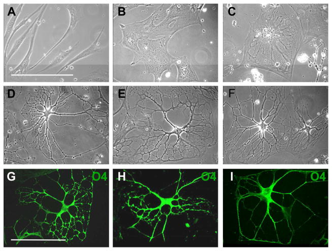
Figure 3.
3D Environment Promotes Further Differentiation of MLPCs. (A–F) Phase contrast images of differentiating MLPCs. (A) The untreated cells displayed typical fibroblast morphology. (B) Cells exhibited flattening and spreading at 24 hrs in 3D environment. (C) Cells at 8 days in the differentiation medium displayed increased flattening. (D, E, F) At 30 days of differentiation, approximately 80% of cells revealed extensive processes. (G–I) Growth factors influenced the development of processes. (G, H) Immunostained cells displaying increased branching and development of processes in presence of bFGF and EGF. (I) Simple processes were observed in absence bFGF and EGF. Scale bars, 100 μm, (40x magnification).

