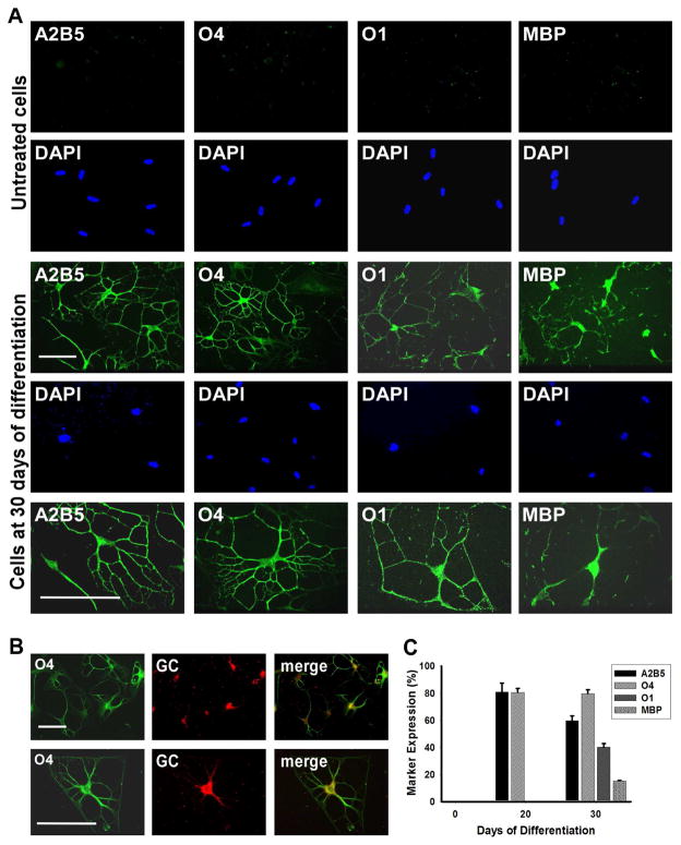
Figure 4.
MLPCs Differentiate into Committed Oligodendrocytes in a 3D Environment. (a) Immunocytochemical analysis of differentiating MLPCs in a 3D environment. The untreated MLPCs indicated negative staining for A2B5, faint staining for O4 and negative staining for O1 galactocerebroside and MBP. At 30 days of differentiation, cells exhibited intensely positive staining for A2B5 and O4. Cells also expressed O1 galactocerebroside and MBP, characteristic of committed oligodendrocytes. Scale bars, 100 μm, (20x and 40x magnification). (b) Co-expression of O4 and galactocerebroside (GC) in the differentiated cells. At 30 days of differentiation, GC was expressed in O4 positive cells. Scale bars, 100 μm, (Row 1, 20x magnification and Row 2, 40x magnification). (c) The progression of differentiation in a 3D environment. At 20 days of differentiation 81.8% of cells expressed oligodendroglial markers A2B5 and 80.6% O4 and were negative for O1and MBP. At 30 days of differentiation, 57.7% of cells stained positively for A2B5, 79.6% for O4, 42.1% for committed oligodendrocyte marker O1 and 15.2% for MBP. Error bars represent the SD.

