Figure 1.
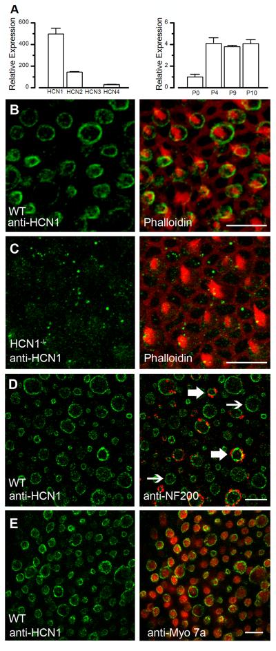
Expression of HCN subunits in the mammalian utricle. A, Quantitative RT-PCR was used to estimate mRNA copy number for each of the four Hcn genes from a pool of 10 utricles collected at postnatal day (P) 8 (left). Data were normalized to Hcn3 mRNA expression. Relative expression of Hcn1 mRNA normalized to 29S and the P0 sample from a pool of 10 utricles collected at four different developmental time points (right). Error bars represent standard error of the mean from three replicates. B, HCN1 in the basolateral hair cell membrane of a mouse utricle (green). Phalloidin (red) is shown in a merged image on the right. The projection of several focal planes through the utricle epithelium reveals that HCN1 is visible in nearly every hair cell basolateral membrane and absent from stereocilia. Scale bar = 10 μm. C, HCN1 staining (green) of an Hcn1-deficient mouse utricle. Phalloidin (red) is shown in a merged image on the right. Scale bar = 10 μm. D, HCN1 in the basolateral hair cell membrane of a mouse utricle (green) counterstained with neurofilmanet-200 (red) to identify afferent terminals. Thin arrows indicate hair cells positive for HCN1 with no visible neurofilament staining, indicating probable type II hair cells. Thick arrows indicate probable type I hair cells positive for HCN1 and surrounded by calyx afferents. Scale bar = 10 μm. E, Confocal image of wild-type mouse utricle hair cells stained with HCN1 (green) and myosin 7a (red). Nearly 90% of hair cells showed HCN1 expression. Scale bar = 10 μm.

