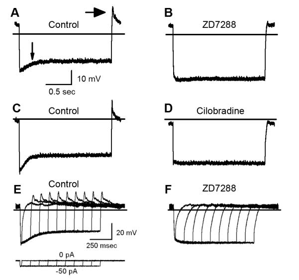
Figure 6.

Hair cell voltage responses. Hair cell voltage responses were measured in current-clamp mode. Two-second current steps of 50 pA or less (panels A-D) were used to hyperpolarize the cell to ~ −90 mV. The horizontal lines indicate −60 mV. The scale bars apply to panels A-D. A, Representative response from a type II hair cell of a wild-type mouse utricle at P8. Following the initial hyperpolarization, the cell depolarized to a steady state level. The depolarizing sag is indicated by a small arrow. When the current was stepped back to 0 pA, the voltage rebounded with a peak near −40 mV (large arrow). B, The same cell shown in A after application of 100 μM ZD7288. C, Other representative wild-type voltage responses before and after (D) application of 10 μM Cilobradine. E, A family of representative voltage responses recorded from a type II hair cell at P7. The responses were evoked by the current protocol shown below. The scale bars apply to panels E-F. F, The same cell shown in E after application of 200 μM ZD7288 and full block of Ih.
