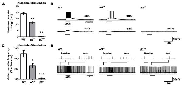
Figure 1. Nicotinic excitability of layer VI pyramidal neurons is reduced in α5-/- and eliminated in β2-/- mice.
A, Stimulation of only nicotinic receptors following blockade of muscarinic receptors by atropine (200 nM) resulted in significant differences in depolarization across all genotypes (P < 0.0001), with markedly less depolarization of α5-/- and β2-/- compared to wild type (WT) neurons (**P < 0.001). B, Sample traces show nicotinic responses in neurons across all genotypes, including the percentages of neurons with suprathreshold (top) and subthreshold (bottom) responses. A smaller proportion of neurons were depolarized to threshold in α5-/- and β2-/- neurons (α5-/-: P < 0.05, β2-/-: P < 0.01) than in WT. C, In neurons already firing action potentials by current injection, nicotinic stimulation affects spiking frequency differently across genotypes (P < 0.0001), with a smaller change in action potential frequency in α5-/- and β2-/- compared to WT neurons (*P < 0.05, **P < 0.001). D, Sample traces show nicotinic responses in neurons depolarized to fire action potentials by current injection. Examples (2 seconds in duration) of baseline and peak responses are shown above each trace.

