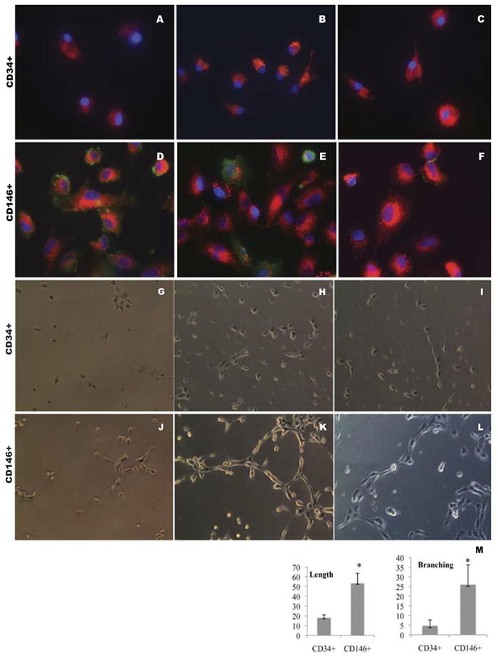
Figure 3.

Effects of mobilized PBMNCs on CD34+ and CD146+ cell differentiation. CD34+ (A-C) and CD146+ (D-F) positive cells were seeded on coverslips and cultured for 3 weeks after which their endothelial specific antigen expression was examined. Green: CD31 (A and D), CD144 (B and E), CD62E (C and F); Red, Dil-LDL; Blue, DAPI. A – F, original magnification, ×600. CD34 (G-I) and CD146 (J-L) positive selected cells were seeded on Matrigel for the tube formation test. After 4 hours (G, J), 18 hours (H, K) and 96 hours (I, L), images were taken using an inverted microscope at 200×. CD146+ progenitor cells formed longer tubes (length) and more interconnections (branching) than CD34 progenitor cells under the same culture conditions. M, mean±SD of the length and branching number (sum of the numbers under each of three random low power fields) for the two cell types, respectively; n=3, * indicates p<0.05 between two groups.
