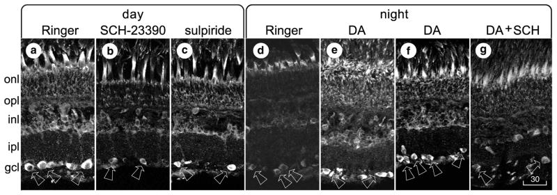
Fig 3.
Receptor profile, using retinas that were dark- or light-adapted, then sectioned, processed, and displayed as in Figure 1. a–c, Pieces of a single light-adapted retina were incubated for 20 min in normal Ringer’s solution (a), 10 μM SCH-23390 (b), or 10 μM sulpiride (c). d, e, Pieces of a single dark-adapted retina were incubated for 30 min in either normal Ringer’s solution (d) or 30 μM dopamine (e). f, g, Pieces of another dark-adapted retina were incubated for 30 min in 30 μM dopamine (f) or for 10 min in 30 μM dopamine, followed by 20 min in 30 μM dopamine plus 10 μM SCH-23390 (g). Magnification is identical in all panels. Scale bar, 30 μm.

