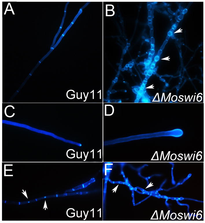
Fig. 2. MoSWI6 deletion results in altered hyphal morphology.
Morphology was determined microscopically after the ΔMoswi6 mutants and the wild type strains were grown for 2 days on CM-overlaid microscope slides. Hyphae were strained with CFW for chitin distribution. Fluorescence indicative of chitin was mainly distributed on the apex of hyphae and septa. (A) and (B) The ΔMoswi6 mutant hyphae showed swelling and became more flexible. (C) and (D) The tips of the ΔMoswi6 mutant hyphae showed expansive growth. (E) and (F) After staining with both DAPI and CFW, no changes were found between the nuclei of the ΔMoswi6 mutants and the wild type strain.

