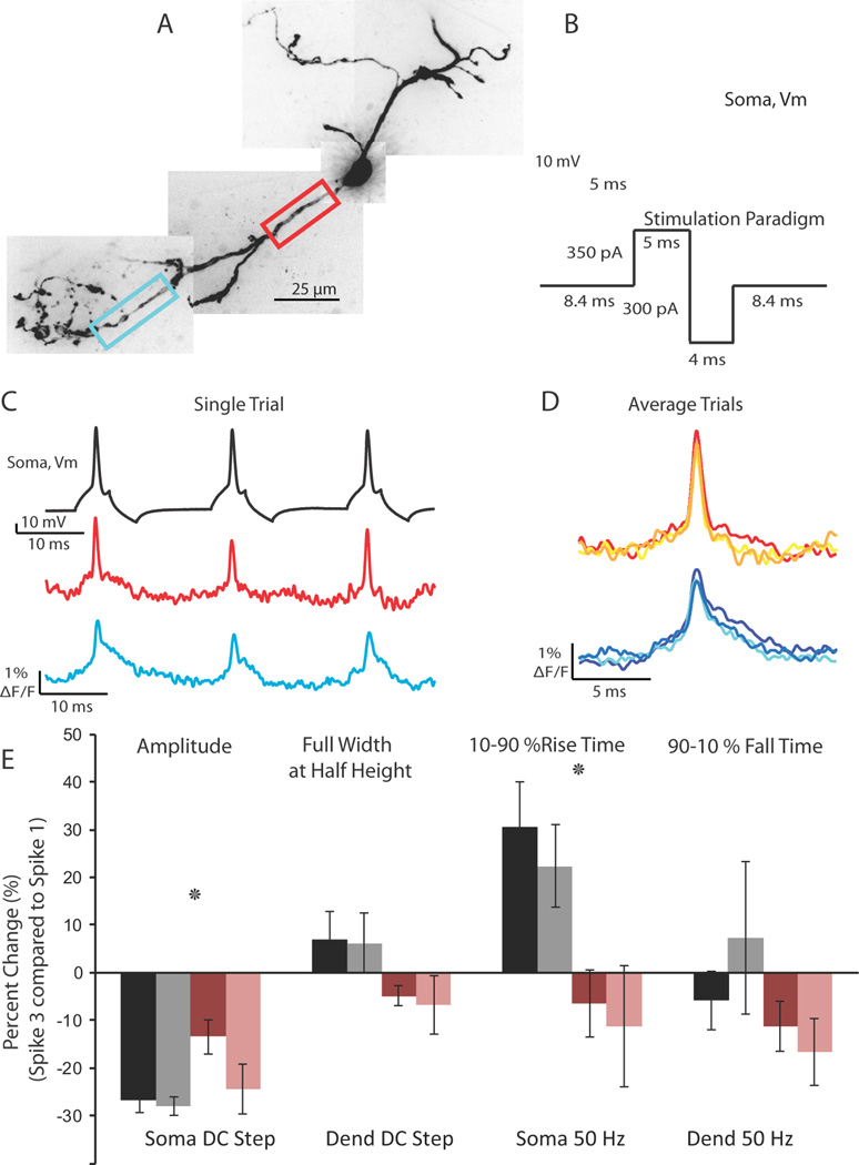
Figure 6.
Action potential spike height decay during trains can be relieved using a 50 Hz depolarizing/hyperpolarizing stimulation protocol. (A) Confocal z-projection of a voltage-sensitive dye filled interneuron. Colored boxes refer to the dendritic regions of interest during imaging. (B) Example of the 50 Hz stimulation protocol used to relieve spike height decay. (C) Single trial example of the multiple action potentials propagating into the dendrite in response to the stimulation protocol in (B). Optical traces were taken from the color boxed regions of interest in (A). Note the lack of extreme action potential height decay. (D) Overlay of 3 action potentials in a 50 Hz train; average of 2 trials. (E) Summary of action potential kinetics (Amplitude, Full Width at Half Height, 10–90 % Rise Time, 90-10 % Fall Time) expressed as percent change between the first and third spike in a train. Black and gray bars refer to changes in action potential waveform at the soma and dendrite, respectively, in response to a 50 ms somatic current injection. Rose and pink bars refer to the changes in action potential waveform at the soma and dendrite, respectively, in response to the 50 Hz stimulation protocol.

