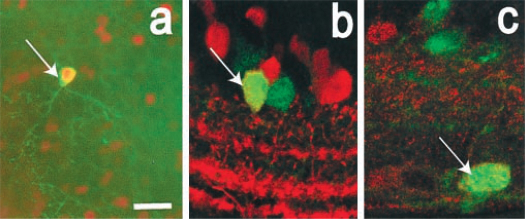
Figure 4.
Colocalization of GFP with marker proteins in retinal amacrine cells. a, Flat-mount view showing tyrosine hydroxylase (TH)-IR (green) and GFP-IR (orange). The arrow indicates a TH+ perikaryon in which GFP-IR is colocalized. b, Vertical section of the retina showing GFP-IR (green), calretinin-IR (red), and colocalization of these two in one neuron (arrow). Note calretinin+ processes in bands within the inner plexiform layer. c, Vertical retinal section showing GFP-IR (green), calbindin-IR (red), and their colocalization in one neuron (arrow). Scale bar (in a) a, 25 µm; b, c, 10 µm.

