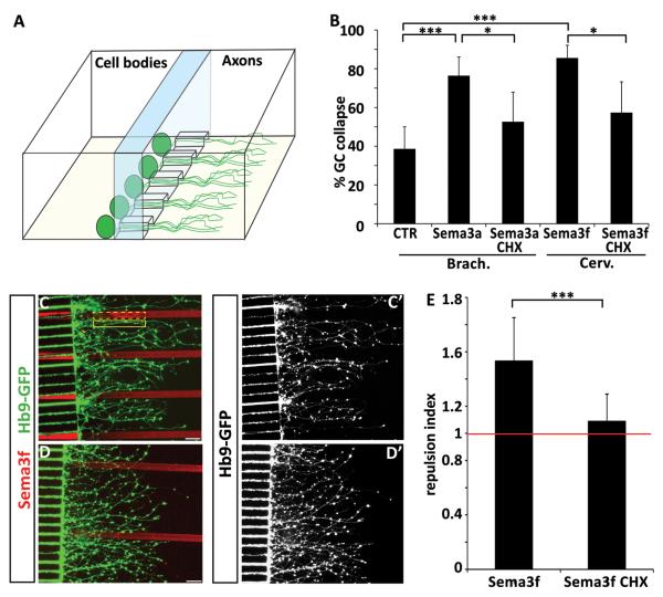
Figure 6. Axonal protein synthesis is required for semaphorin-mediated collapse and guidance.
(A) Graphic representation of a microfluidic chamber. Differentiated embryoid bodies are plated in one compartment of the chamber. Axons grow through microchannels and enter the “axonal compartment” – see also Shi et al, 2010. (B) Sema3a induced collapse of brachio-thoracic motor axons and Sema3f induced collapse of cervical motor axons are attenuated when cycloheximide is applied to the axonal compartment of the microfluidic chamber. (C-C’) Micropatterned Sema3f stripes (red) are avoided by cervical motor axons, but treatment of axons with cycloheximide attenuates Sema3f avoidance (D-D’) (scale bars : 40 μm). (E) The repulsion index is calculated as a ratio of GFP intensity in two adjacent regions of interest of identical size – one covering the laminin stripe (solid yellow line in C) and the second covering the Sema3f stripe (dash yellow line in C). The repulsion mediated by Sema3f (index 1.53) is significantly (t-test t-test p< 0.001) attenuated by cycloheximide treatment (index 1.088) (n=16 stripes/condition).

