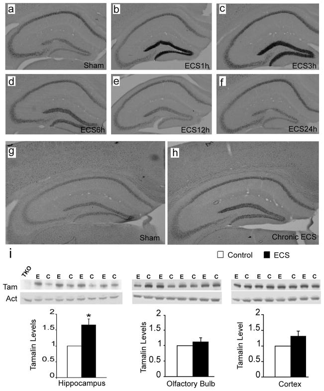
Figure 2. Hippocampal tamalin mRNA and protein are up-regulated in response to ECS.
Representative tamalin in situ hybridization analysis of hippocampus from mice after single (a–f) or chronic (g, h) ECS. Sham-treated animals show the basal level of tamalin expression (a, g). Tamalin mRNA is up regulated at 1 hour (b) and peaks at 3 hours (c) from ECS. Beginning at 6 hours (d) tamalin is down regulated and returns to the pre-ECS level by 12 (e) and 24 hours (f). Note that tamalin mRNA expression remains up-regulated at 24 hours after chronic ECS (h) as compared to sham-treated controls (g). n=4 for each group. Expression of tamalin protein after cronic ECS (i). Animals received one ECS a day for 5 consecutive days and were sacrificed 24 hrs after the last treatment. Western blot analysis of hippocampus (left panel), olfactory bulb (central panel) and cortex (right panel) from ECS (E)- or sham (C)-treated WT animals using an anti-tamalin antibody (Tam); b-actin was used as loading control (Act) and a hippocampus lysate from a tamalin KO mouse was used as negative control (TKO). The quantification of tamalin protein levels is shown at the bottom. Note the selective tamalin up regulation in the hippocampus. n = 4 animal. P < 0.05 students ‘t’ test.

