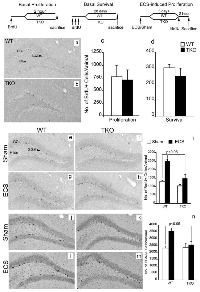
Figure 4. Tamalin deficiency blocks ECS-induced proliferation of adult hippocampal progenitors.
BrdU staining of representative hippocampal sections from WT (a) and tamalin KO (b) mice showing BrdU+ hippocampal progenitors in the subgranula zone (SGZ, arrowhead) of the DG, 2h post BrdU injection. (c, d): Quantification of BrdU+ cells in the SGZ of the hippocampus showing no changes in basal progenitors proliferation (c) or survival (d) in WT and Tamalin KO mice. The data analyzed by student’s t test represent the mean ± SEM; n = 4–5 per group. (e–h): Representative BrdU immunohistochemistry images from sham (e, f) or ECS-treated (g, h) WT (e, g) and tamalin KO (f, h) animals. Note the mild, non significant, increase of ECS-induced BrdU+ proliferating progenitors in the SGZ of the DG of TKO mice compared to the robust increased in the WT animals (i). (j–m) Representative photomicrographs of PCNA+ cells as in (e–h). Note the significant increase in the number of PCNA+ cells only in the ECS-treated WT animals (n). Data in (i, n) was analyzed by two-way ANOVA followed by post hoc Bonferroni test and represent the Mean ± SEM. n = 8–10 per group. Two-way ANOVA revealed a significant ECS and genotype interaction for data presented in (i) (F(1, 14)= 6.53, p <0.05) and (n) (F(1, 32)= 5.87, p <0.05). The schedules of BrdU injection and analysis for the basal proliferation (c), survival (d) and ECS-induced proliferation (i, n) are depicted at the top. GCL, granular cell layer.

