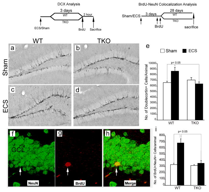
Figure 5. Tamalin is required for ECS-induced hippocampal neurogenesis.
(a–d): Representative immunohistochemistry images showing doublecortin-positive (DCX+; a marker for immature neurons) cells in the dentate gyrus of WT (a, c) and Tamalin KO (b, d) animals subjected to sham (a, b) or ECS treatment (c, d). (e): Quantitative analysis of DCX+ cells showing an increase in the number of immature neurons caused by ECS in WT but not in tamalin KO animals. n= 6–8 animals per group. (f–h): Confocal z-stack representative images showing co-localization of BrdU+ cells with NeuN, a marker for mature neurons. WT and Tamalin KO animals were subjected to a single sham or ECS treatment and they were injected with BrdU (200mg/kg i.p.) three days later. (i): Quantitative analysis showing the increase in the number of BrdU/NeuN double positive cells caused by ECS in WT animals but not in tamalin KO mice. n = 4–7 animals per group. Data represent the Mean ± SEM analyzed by two way ANOVA followed by Bonferroni posthoc test. Two-way ANOVA demonstrated a significant ECS-genotype interaction in both the DCX (e) (F(1, 24)= 8.01, p <0.05) and NeuN/BrdU (i) (F(1, 19)= 8.77, p <0.05) analysis. The schedules of BrdU injection and DCX or BrdU/NeuN analysis are depicted at the top.

