
Figure 4.
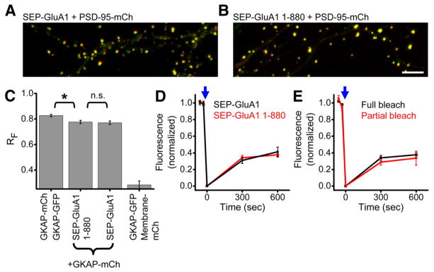
GluA1 PDZ ligand deletion does not alter receptor positioning or mobility. A, B, Dendrite regions from neurons expressing either full-length SEP-GluA1 (A) or SEP-GluA1 1–880, lacking the PDZ ligand (B), and PSD-95-mCh. Scale bar, 10 μm. C, Mean RF for neurons expressing the indicated constructs. (n =4, 9, 12, 6 neurons in the order shown). Statistics: Kruskal–Wallis ANOVA (p ≪ 0.001) post hoc pairwise comparisons by Mann–Whitney U test, *p <0.05. D, E, SEP-GluA1 fluorescence recovery after either full or partial synapse photobleaching. Experiments were interleaved in neurons coexpressing either SEP-GluA1 or SEP-GluA1 1–880. n =9–31 synapses from 5–10 neurons.