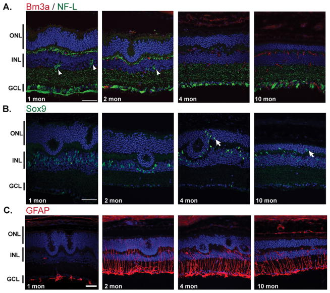
Figure 3. Altered cell types in 1, 2, 4 and 10-mon Nrl−/− retina.
(A) Ganglion cells were immunolabeled with anti-Brn3a (red) and anti NF-L (green) and showed presence of displaced ganglion cells in the INL of young animal (1 and 2-mon old). These were not present in 4 and 10-mon old animals. (B) Muller cells, immunolabeled with anti-Sox9 antibody (green), migrated first around the remaining pseudorosettes (at 4-mon) then in the entire outer nuclear layer (10-mon). (C) GFAP immunolabeling (red) showed strong activation of Muller cells at 2, 4 and 10-mon of age.

