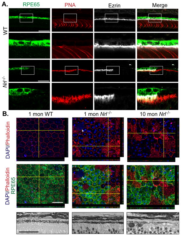
Figure 5. Changes in RPE in Nrl−/− retina.
(A) Immunolabeling on vibratome sections of RPE with anti-RPE65 (green), cones with PNA (red) and RPE apical side with anti-Ezrin (grey) on 4-month old WT and Nrl−/− mouse retina showed weaker RPE65 expression and absence of ezrin in front of cone photoreceptors in Nrl−/− mice compared to WT mice. Scale bar: 40μm (B) Top Panel: RPE-sclerochoroidal whole mount of 1 month WT mice and 1- and 10-mon old Nrl−/− mice after immunolabeling of RPE cellular outlines stained with Phalloidin (red) and anti-RPE65 (green). Compared to RPE from WT mice, RPE from Nrl−/− mice showed abnormalities in their junctions with presence of large patches positive for phalloidin. Nuclei are visualized by DAPI staining. Arrowhead indicates apoptotic bodies. If in WT mice, RPE65 showed homogenous expression all RPE cells, in contrast RPE65 expressions were absent in a large number of RPE cells and even among the cells with phalloidin staining in their cell body. Bottom Panel: High magnification of methacrylate sections followed by H&E staining showed a loss of RPE cells in aged (10-mon) Nrl−/− retina. Scale bar = 20μm.

