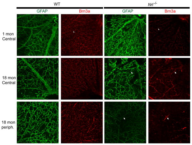
Figure 7. Age-associated alterations of the superficial retinal vasculature and ganglion cell death in Nrl−/− retina.
Immunohistochemistry on whole-flat mount retina with anti-GFAP (green) and anti-Brn3a (red) antibodies labeling astrocytes, activated Muller cells and ganglion cells, respectively. GFAP-positive cells aligning blood vessels on the ganglion cell layer side showed profound disorganization of the retinal vasculature, with large areas with no staining in 18-mon old Nrl−/− mice compared to WT control. Presence of ganglion cells was assessed with anti-Brn3a immunolabeling. Nrl−/− retina did not show high level of Brn3a expression at all time points. Loss of ganglion cells occurred in 18-mon old Nrl−/− mice but was more pronounced at the periphery compared to central retina.

