Figure 3.
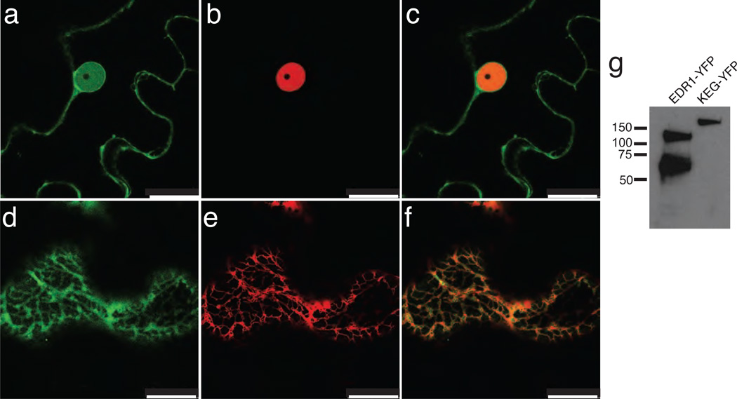
Subcellular localization of EDR1. Panels a–c, EDR1-sYFP and GCN5-mCherry were transiently co-expressed in N. benthamiana leaves and imaged using confocal laser scanning microscopy. Panel a, EDR1-sYFP (a single optical section taken through the nucleus of an epidermal cell). Panel b, GCN5-mCherry expressed in the same cell. Panel c, overlay of panels a and b. Panels d–f, EDR1-sYFP and an mCherry ER marker (see Methods) were transiently co-expressed in N. benthamiana. d, EDR1-sYFP (a single optical section taken through the cell cortex of an epidermal cell); e, mCherry-HDEL, f, overlay of d and e. Panel g, immunoblot of EDR1-sYFP extracted from N. benthamiana leaves. KEG-sYFP is an unrelated YFP fusion protein included to show specificity of the antibody. Scale bar is 25 µm.

