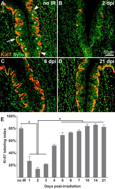
Figure 1. Irradiation targets actively cycling cells.
Proliferative cells in taste epithelium are marked by Ki67-IR (red) and nuclei are visualized with Sytox (green). Compared with controls (A), the number of basal proliferative cells is significantly reduced at 1–3 dpi (B, E), then returns to and is maintained at control levels from 5 to 21 dpi (C: 6dpi; D: 21 dpi). White asterisks indicate taste buds. White arrows indicate active cycling cells in basal epithelium. Total basal epithelial cell nuclei and Ki67-IR cells were counted between the arrowheads in each trench. E: Bar graph of the proportion of Ki67-IR basal epithelial cells at progressive time points after irradiation (Mean +/− SD; n = 3–5 mice for each time; one-way ANOVA, Tukey post hoc test, * p < 0.01.

