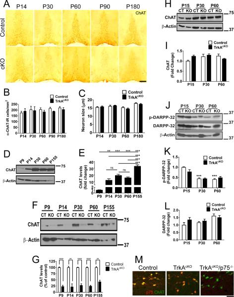
Figure 5.
TrkA loss affects ChAT expression in BFCNs, but not striatal neurons. A, Neurons in the medial septum of P14, 30, 60, 90, 180 of control and TrkAcKO mice stained with anti-ChAT antibody. Neurons of mutant mice exhibited decreased ChAT staining compared to control. B, Quantitative analysis of ChAT-IR neuronal cell numbers of control and TrkAcKO mice. n=3–4 mice per genotype. C, Evaluation of control and TrkAcKO ChAT-IR neuronal cell size. n=35 cells per genotype analyzed among 3 mice. D,E, ChAT protein levels in BF of control mice at different ages. n=4. F,G, Quantitative western blot of ChAT levels in BF. A reduction in ChAT expression in the TrkAcKO mice compared to WT mice is already seen at P9 and is sustained throughout cholinergic development. n=4 mice per genotype at P9, P14; n=7–8 at P30, P60; n=3 at P155. (H–L) Analysis of cholinergic signaling in striatum of TrkAcKO mice. Whole tissue lysates were prepared from striatum of either control or mutant animals at various ages and subjected to western blotting using the indicated antibodies. (H,I) Total protein levels of ChAT remain unaffected in mutant animals. n=4 mice per genotype. (J–L) Unaltered DARPP-32 signaling in striatum of TrkAcKO mice. ***, compared to P15-control; n=6 mice per genotype. M, Neurons in the medial septum of P30 control, TrkAcKO and TrkAcKO/ p75−/− double mutant mice stained with anti-ChAT and anti-p75 antibodies. Although there is complete absence of NGF signaling in double mutant mice (right panel), there is no significant change in BFCN morphology or ChAT expression compared to TrkAcKO littermates. Significance values: E, G, K, *p<0.05, **p<0.01, ***p<0.001, ****p<0.0001. Scale bars: A, 500 μm; M, 50 μm.

