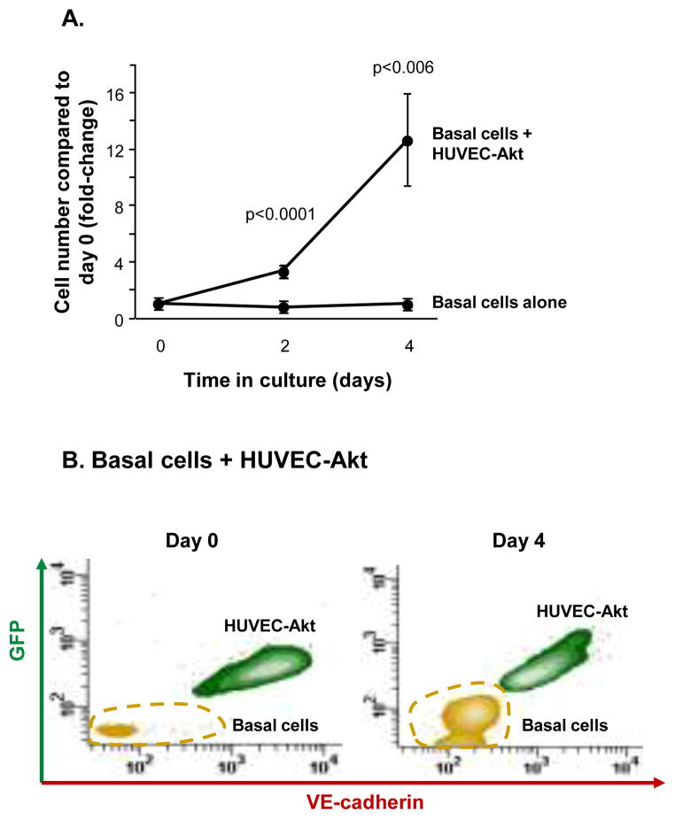
Figure 7.
Endothelial cells support the growth of airway basal cells in the absence of growth factors. A. Human airway basal cells were cultured alone or in co-culture with Akt-activated human umbilical vein endothelial cells (HUVEC-Akt) in cytokine- and serum-free conditions. At the desired time points, cells were harvested and the GFP-labeled HUVEC-Akt cells was determined as the GFP+VE-cadherin+ population by flow cytometric analysis, and the GFP−VE-cadherin− population quantified as expanded basal cells. Data shown is the average of 4 independent experiments. B. Representative flow cytometric analysis of human airway basal cell and HUVEC-Akt populations at day 0 and day 4 of co-culture. HUVEC-Akt cells were determined as the GFP+VE-cadherin+ population, and the GFP−VE-cadherin− population quantified as expanded basal cells.

