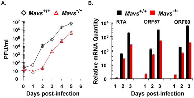
Figure 2.
γHV68 lytic replication kinetics in mouse embryonic fibroblasts (MEFs). The lytic replication of γHV68 on Mavs+/+ and Mavs−/− MEFs was assessed by multi-step growth curves (A) and quantitative real-time PCR (B). For both experiments, equal number of MEFs and amount of γHV68 were used for viral infection at a multiplicity-of-infection (MOI) of 0.01. (A) Cells and supernatants were harvested at indicated time points and subject to a plaque assay to determine viral titers. (B) Total RNA was extracted from γHV68-infected MEFs and analyzed by quantitative real-time PCR with primers specific for selected lytic transcripts (RTA, ORF57, and ORF60).

