
Figure 1. An antibody to cone arrestin, 7G6, labels primate cones.
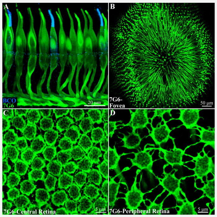
A. Cones in the macaque retinal sections were labeled with 7G6, a cone arrestin antibody, shown in green. Cones were labeled in their entirety with particularly bright staining of the outer segments and pedicles or synaptic terminals. Red/green cones cannot be distinguished but double labeling with an antibody against blue cone opsin (blue) shows that blue cones are also stained by 7G6. 8 × 0.5 μm optical sections
B. A wholemount view of the macaque fovea shows cones labeled with the 7G6 antibody. The axons of the foveal cones are displaced radially away from the foveal pit, forming Henle’s fiber layer, and terminating in a ring of cone pedicles around the fovea. 20x objective, 9 × 1 μm sections.
C. Central primate retina, focus at the level of the outer plexiform layer. Cone pedicles, labeled with 7G6, are densely packed, often in contact. They also extend short processes, called telodendria, to contact adjacent pedicles. The rim of each cone pedicle is densely stained but there are many holes in the base of the cone pedicle that correspond to synaptic invaginations (see below). 5 × 0.4 μm optical sections.
D. In peripheral primate retina, the cone density decreases, the cone pedicles are widely spaced and the telodendria lengthen to compensate. 5 × 0.4 μm optical sections.