
Figure 2. 7G6 antibodies outline synaptic invagination sites.
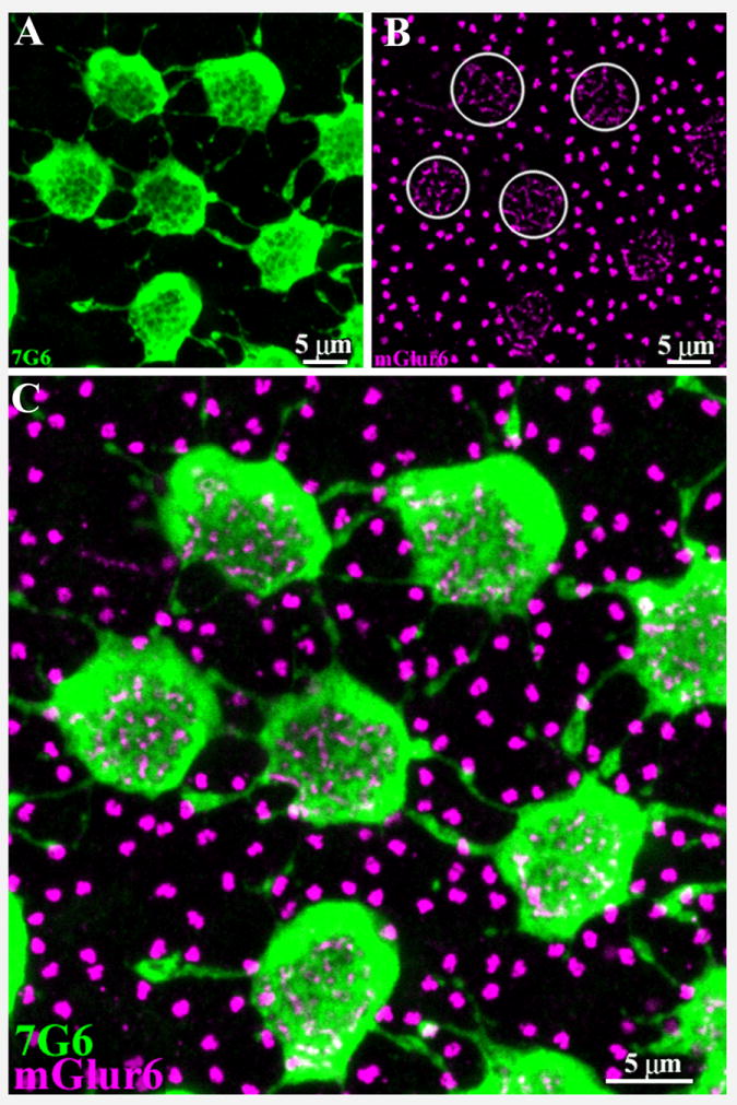
A. A high-resolution image of 7G6-labeled cones shows several cone pedicles interconnected by telodendria. Some cone telodendria appear to have enlarged endings in peripheral retina. As in figure 1, there are holes in the central base of each cone pedicle.
B. The tips of ON cone bipolar cells and rod bipolar cells contain mGluR6. Antibodies against mGluR6 (magenta) stain two distinct structures in the outer plexiform layer. The tips of ON cone bipolar cells appear as clusters of small mGluR6 labeled processes, which invaginate the cone pedicle (several circled). The tips of rod bipolar cells appear as brightly stained doublets that occupy the space between cone pedicles. These also indicate the positions of unlabeled rod spherules.
C. Double label image shows that the holes in the center of 7G6-labeled cone pedicles colocalize with the mGluR6 signals of ON cone bipolar cells. This indicates that the dendritic endings of ON cone bipolar cells enter each hole in the base of the cone pedicle. Therefore, the holes represent synaptic invagination sites and indicate that the level of focus is at the very base of the cone pedicle. 5 × 0.5 μm optical sections.