
Figure 3. Cx36 labeling occurs both between and underneath cone pedicles.
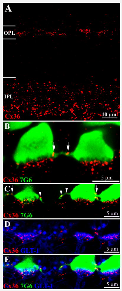
A. A cross section of macaque retina shows Cx36 labeling in both plexiform layers. The prominent Cx36 labeling in the inner IPL is mainly associated with AII amacrine cells. In comparison, the Cx36 labeling in the OPL is fainter; the plaques are smaller and form clusters.
B. Double labeling shows that Cx36 plaques occur in two locations: on fine processes between 7G6 labeled cone pedicles (arrows) and in prominent clusters underneath each cone pedicle. The Cx36 plaques underneath the cone pedicle are distinct from and do not colocalize with the base of the cone pedicle.
C., D. E. Triple label images from vertical sections of macaque retina. C. Cone pedicles are stained with 7G6 (blue). Cx36 plaques (red) occur at cone pedicle contacts (arrows) and on fine processes leaving the cone pedicle base (arrowheads). D. Certain OFF bipolar cells, including OFF midget bipolar cells and DB2, are labeled with antibodies against the glutamate transporter GLT-1 (green). The OFF bipolar dendrites terminate in flat tops, apposed to the base of the cone pedicle. Most of the Cx36 labeling underneath the cone pedicles is colocalized with the OFF bipolar dendrites. E. Triple label image shows that the Cx36 plaques below each cone pedicle are colocalized with GLT-1 stained OFF bipolar cells and not with the cone pedicle. All panels, 6 × 0.4 μm optical sections.