
Figure 8. Cone pedicles make potential Cx36 gap junctions with most surrounding rod spherules.
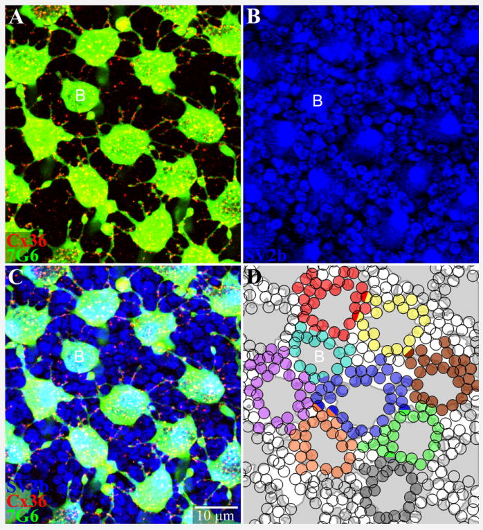
A. A mosaic of cone pedicles stained with 7G6 (green). Many telodendria, long and short, are decorated with Cx36 plaques (red). A single small blue cone pedicle is marked with a letter B. B. Same field showing rod spherules, which are round with a dark central spot stained for SV2b (blue). Cone pedicles are larger polygonal structures. C. Triple-label image shows Cx36 contacts with many neighboring rods and cones. D. A reconstruction of part of the cone pedicle mosaic. The cone pedicles were outlined and colored grey. Each rod spherule receiving a Cx36 telodendrial contact was shaded with one color indicating contacts from a single cone pedicle. A few rods receiving Cx36 contacts from two different cones were coded as hemispheres of two colors, including at least two rod spherules with blue input. The small telodendria extend for 2-3 rows of rod spherules around each cone pedicle. Almost every rod spherule receives potential gap junction contacts. Red/green cone pedicles contacted 20-30 adjacent rods. A single blue cone pedicle (B) made potential contacts with 16 rod spherules. 6 × 0.3 μm optical sections.