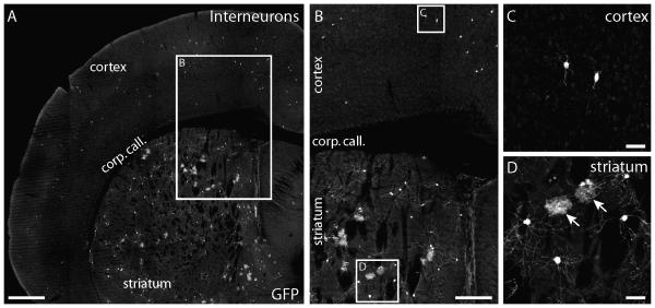
Figure 2. Interneurons in cortex are generated independently of cortical astrocytes.
Approximately 5% of adult TFC.09 x Z/EG mice exhibited widely distributed GFP+ cells within cortex that were not arranged in columns. In these mice, GFP+ cells were distributed sparsely in layers 2 through 6 of neocortex through the entire anteroposterior and mediolateral axes. The GFP+ cells in cortex of these mice displayed the characteristic morphologies of interneurons, with fine local processes. The cortices of these mice did not contain labeled astrocytes. TFC.09 x Z/EG mice with interneurons in cortex always had GFP+ cells broadly distributed through their ipsilateral striatum. In mice with GFP+ interneurons in cortex, the striatum contained both GFP+ neurons and astrocytes. In the striata of these mice, GFP+ neurons and astrocytes had the characteristic morphologies of medium spiny interneurons and protoplasmic astrocytes, respectively. 500 μm (A), 250 μm (B), and 50 μm (C, D)

