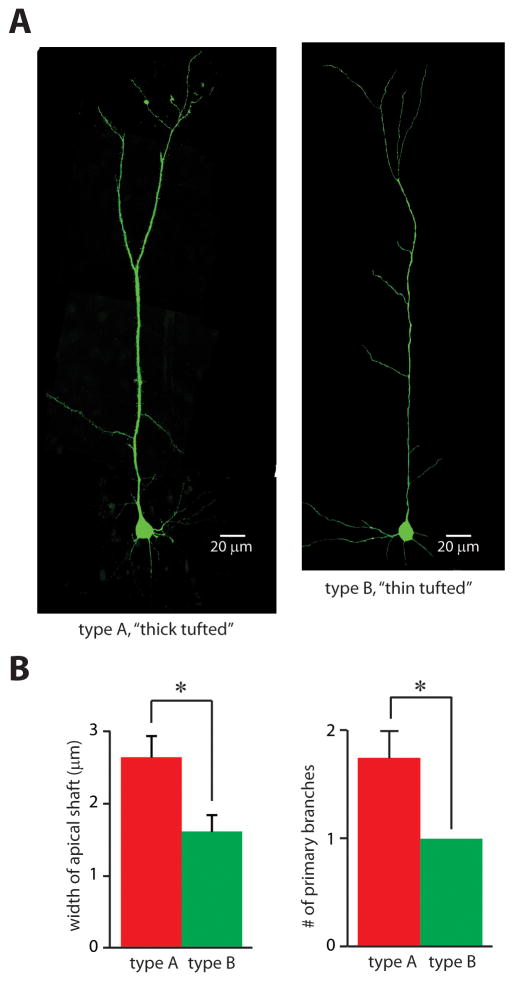
FIGURE 2. Type A and B pyramidal neurons have different morphologies.
(A) Confocal images of representative neurons in which the amount of h-current falls either above (“type A,” left image) or below (“type B,” right image) the threshold in Fig. 1C. (B) Type A and B neurons differ in the widths of the shafts of their apical dendrites (left) and in the number of primary branches of their apical dendrites (right) (n = 4 neurons in each group). *p < 0.05.

