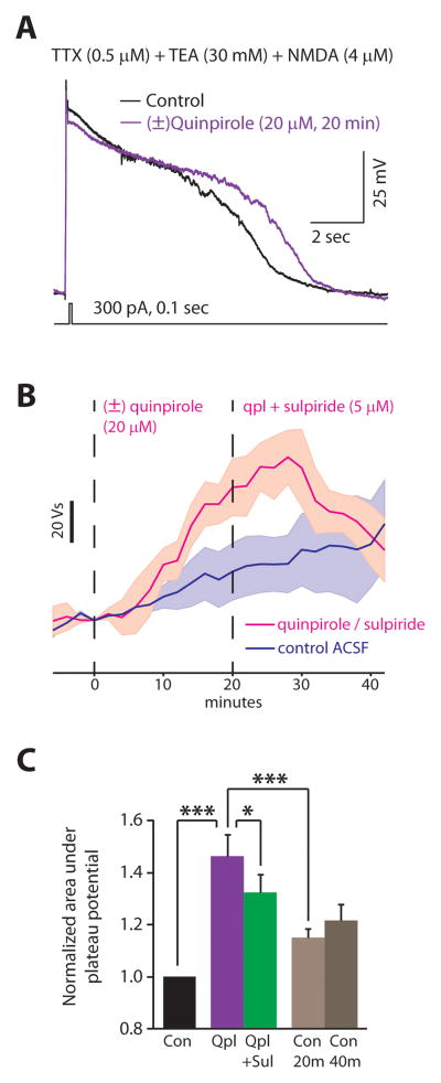
FIGURE 7. Quinpirole reversibly prolongs calcium-dependent plateau potentials.
(A) A short current pulse elicits a brief Ca2+ spike that is followed by a prolonged plateau potential in a type A neurons after application of TTX and TEA. Each experiment recorded plateau potentials in control conditions, then while applying (±)quinpirole (20 μM), and finally while applying quinpirole + sulpiride (5 μM). (B) We quantified the size of plateau potentials by measuring the area under the voltage trace. The average size of the plateau potentials are shown as a function of time (n=4 cells in each condition). For the magenta trace, t=0 represents the beginning of quinpirole application, and quinpirole and sulpiride were both applied after t=20 min. The dark blue trace represents recordings in control ACSF. Shaded regions represent ± 1 S.E.M. (C) Summary data for the size of plateau potentials in each condition. Each bar represents data collected from 5 min before until 5 min after the end of drug application, or corresponding timepoints during recordings in control ACSF. We measured plateau potentials every 5 min during this period and used repeated measures ANOVA and corrections for multiple comparisons to assess statistical significance. * = p < 0.05, *** = p < 0.001.

