Figure 1.
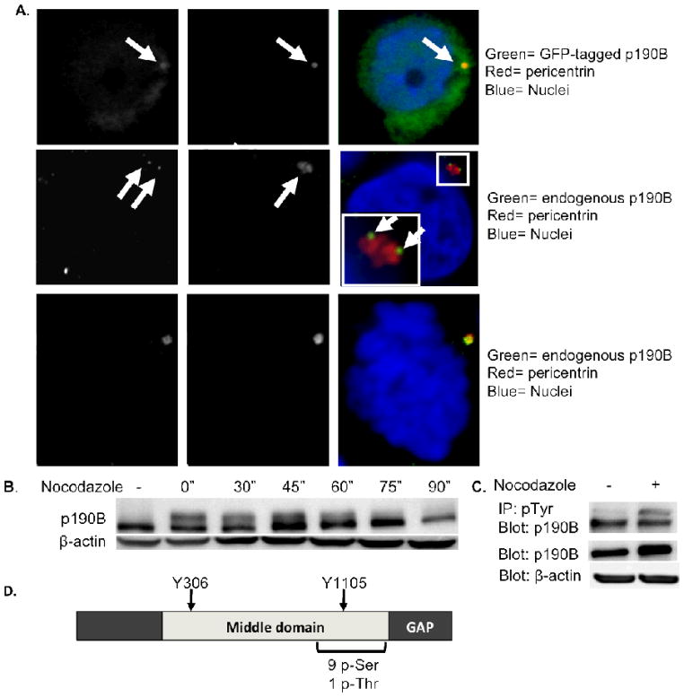
P190B RhoGAP localizes to centrosomes and is phosphorylated during mitosis. Confocal images are shown demonstrating co-localization of GFP-tagged p190B and endogenous p190B protein with pericentrin in interphase MCF-7 cells, as well as co-localization of endogenous p190B with pericentrin in a mitotic MCF-7 cell (A). Western blot of nocodazole synchronized MCF-7 cells reveals a slower migrating form of p190B during mitosis. β-Actin is shown as a loading control (B). Western blot of immunoprecipitated pTyr proteins from asynchronous and synchronized MCF-7 cell lysates probed with an anti-p190B antibody demonstrating that p190B is differentially phosphorylated on Tyr residues during mitosis (C). P190B levels are comparable in asynchronous and synchronized lysates as shown by a Western blot loaded with 10% input and probed with p190B antibody. β-Actin is shown as a loading control. Diagram depicts p190B phosphorylation sites (D). P190B is phosphorylated on 9 Ser residues and 1 Thr residue during mitosis.

