
Figure 2.
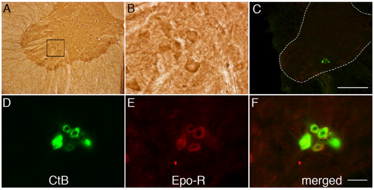
Representative images of EPO receptor (EPO-R) immunostaining in C4 phrenic motor neurons. (A) DAB staining revealed EPO-R expression in large, putative phrenic motor neurons (black box) and interneurons. (B) Higher magnification of black bock from panel A. (C) CtB labeled phrenic motor neurons (green cells in C4 ventral horn). Scale bar, 400μm. (D-F) EPO-R (E) is expressed in CtB labeled phrenic motor neurons (D; see merged image in F) and the surrounding neuropil. Sections were incubated without primary or secondary antibody as negative controls. Scale bar in F is 50μm.