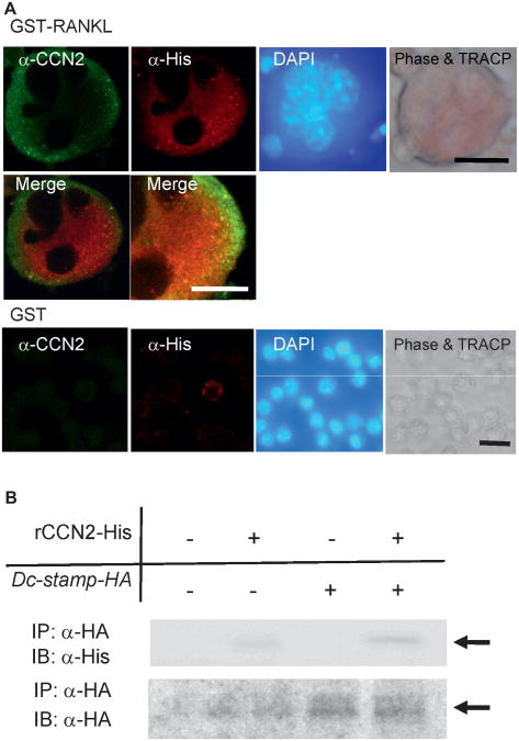
Fig. 4.
Colocalization and interaction of CCN2 with DC-STAMP in osteoclast-like cells. (A) Immunofluorescence analysis of CCN2 and DC-STAMP expression on RAW264.7 cells treated with GST-RANKL. RAW264.7 cells were inoculated at a density of 1 × 10⁴/cm2 into a 4-well chamber slide, and the next day the medium was removed and infected with retroviruses, having pMGF-Dc-stamp-His, containing 8 μg/mL of polybrene (hexadimethrine bromide). After 8 hours, the retrovirus solution was replaced with fresh α-MEM containing 10% serum with GST-RANKL or GST, and the cells were cultured for 7 days. Thereafter, RAW264.7 cells that had differentiated into multinucleated giant cells were analyzed by laser-scanning confocal microscopy. The same field was viewed by phase-contrast microscopy, nuclear staining with DAPI, and TRACP staining. CCN2 showed cell surface distribution (green), and DC-STAMP showed cell surface plus cytoplasmic distribution (red). In the merged image, colocalization of the two molecules was observed, and the highermagnification image showed the colocalization of two molecules more clearly. On the other hand, CCN2 and DC-STAMP was at very low levels in RAW cells treated with GST. The bar represents 25 μm and 10μm (high-magnification image). (B) Western blot analysis of CCN2 binding to DC-STAMP immunoprecipitated with anti-HA antibody. RAW264.7 cells were inoculated at a density of 1 × 10⁵ /well into a 6-well multiwell plate, and the next day the medium was replaced with serum-free medium. Then the cells were transfected with a DC-STAMP expression plasmid containing an HA tag by using FuGENE6. After 18 hours, the medium was replaced with fresh α-MEM containing 10% serum with GST-RANKL or GST, and the cells were cultured for 2 days. Thereafter, the cell lysates were collected and incubated without or with 1 μg rCCN2 tagged with His at 4°C for 2 hours. Then immunoprecipitaion was performed with Ezview Red anti-HA affinity gel (Sigma) at 4°C. After 1 hour, precipitates were subjected to SDS-PAGE and Western blotting with anti-His antibody (upper panel). The arrow indicates the signal for CCN2. In the lower panel, the same membrane was reacted with anti-HA antibody. The arrow indicates the signal for DC-STAMP-HA.

