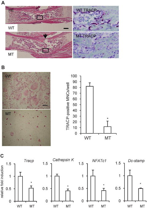
Fig. 6.
Impaired formation of TRACP+ multinucleated cells generated by M-CSF- and RANKL-treated fetal liver cells from Ccn2 null mice. (A) Hematoxylineosin (H-E) staining of radii from wild-type and Ccn2 null mice (left panel) and TRACP staining in the box of H-E staining (right panel) on embryonic day 18.5. The bar represents 200 μm. The number of TRACP+ cells was decreased in Ccn2 mutants compared with wild-type mice. The arrow indicates the concave surface of the kink in Ccn2 mutants. (B) To investigate the effect of CCN2 on osteoclastogenesis, we isolated fetal liver cells from Ccn2 null mice on embryonic day 14.5 and treated them with M-CSF (10 ng/mL) for 3 days. The M-CSF-treated adherent cells were treated with M-CSF and GST-RANKL for 7 to 10 days, fixed, and stained for TRACP. (Left panel) The cells from wild-type mice (WT) treated with M-CSF and GST-RANKL differentiated into TRACP+ multinucleated cells (MNCs), whereas the formation of TRACP+ MNCs from Ccn2 null mice (MT) was impaired. The bar represents 50 μm. (Rightpanel) The graph shows quantification of TRACP+ MNCs with more than three nuclei. Data are the means ± SDs (n = 4). The asterisk indicates a significant difference from WT (p<.01). (C) The expression of osteoclast marker genes was determined by real-time RT-PCR. Real-time RT-PCR analysis was performed as described under “Materials and Methods.” Data are presented as means and SDs for wild type (n = 3) and Ccn2 null (n = 4) mice. The asterisk indicates a significant difference from WT (p < .05).

