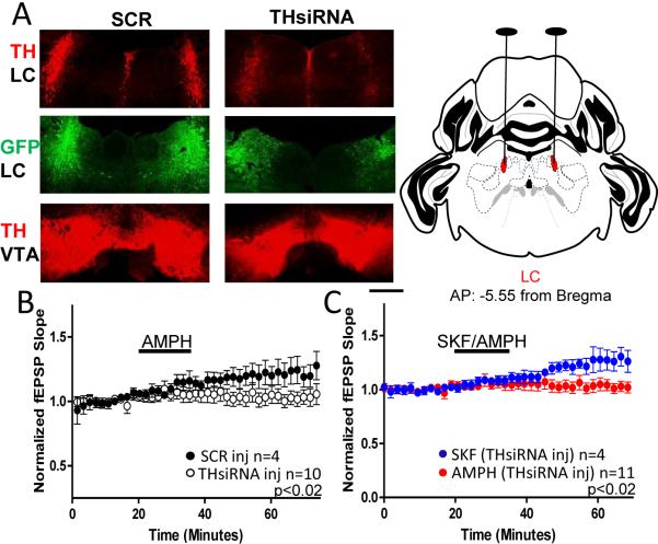
Figure 3.
Loss of TH in the LC is sufficient to prevent the D1 mediated increase in glutamate transmission with AMPH stimulation. (A) Left, Top: Confocal images from an AAV-THsiRNA and an AAV-SCR injected mice acquired at 5× represent mean fluorescence from each group. TH staining is observed in the LC of AAV-SCR injected mice but is diminished in mice injected with AAV-THsiRNA. Bubble in left panel removed in photoshop. Left, Middle: GFP staining shows the precise targeting of virus to the LC. Left, Bottom: TH staining is observed in the VTA region from mice injected with either AAV-THsiRNA or AAV-SCR in the LC. Bubble in right panel removed in photoshop. Magnification bar indicates 0.5mm.Right, Top: Coronal drawing depicts bilateral location of LC and surgical targeting adapted from reference mouse brain atlas: Allen Institute for Brain Sciences. (B) Plot shows AMPH increases glutamate transmission in slices from AAV-SCR injected but not AAV-THsiRNA injected mice (THsiRNA: 100±4%; n=10; SCR: 120±8%; n=5; p<0.02). (C) Plot shows SKF treatment increases glutamate transmission in slices from AAV-THsiRNA injected mice (AAV-THsiRNA SKF:126±8%; n=5; AMPH vs. SKF p<0.02). AMPH experiments are replotted for comparison.. Error bars represent SEM.

