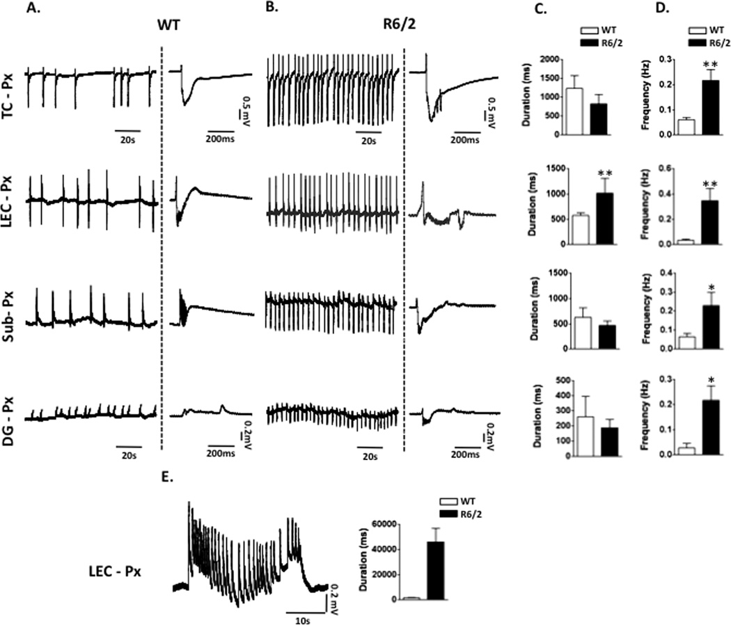
Figure 4. Prolonged epileptiform discharges in LEC in slices from R6/2 mice after GABAA receptor blockade.
A. Shown are spontaneous LFPs after 30 min bath application of 50 µM picrotoxin in the regions indicated in WT mice, to the right of each trace there is a characteristic LFP induced by blocking GABAA. B. Changes induced by picrotoxin in R6/2 mice differ significantly from WT mice, shown are representative recordings and close up views of single events in the different regions. C. and D. Comparisons of LFPs duration and frequencies (mean values and SD) in the different regions between WT and R6/2 mice. E. Example of an epileptiform discharge induced by 50 µM picrotoxin in LEC in an R6/2 mouse, to the right are plots of the LFP ≥ 1.5 sec in duration in this region.
St= striatum, TC=temporal cortex, PMC=primary motor cortex, PFC=prefrontal cortex
* denotes p value <0.05, and ** p value < 0.01

