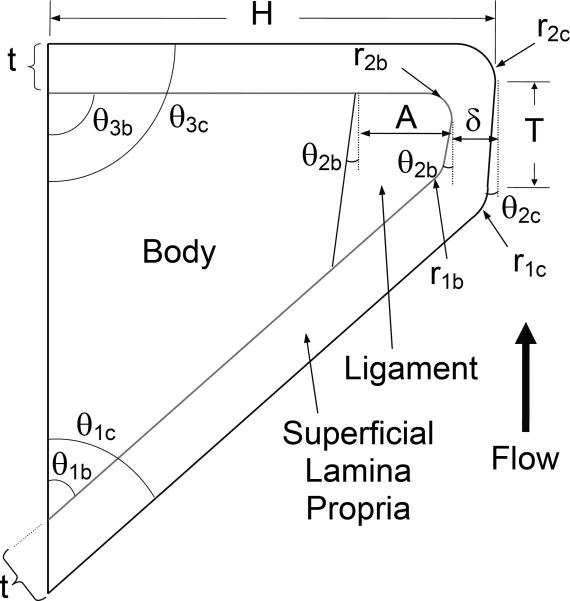
Figure 2.
Synthetic vocal fold model cross section. Distinct body, superficial lamina propria, ligament, and epithelium layers are shown. Parameters define vocal fold model geometry. This figure is scaled for clear representation of geometric definitions. Application of the parameter values given in Table 1 will result in a slightly different shape than what is shown here.

