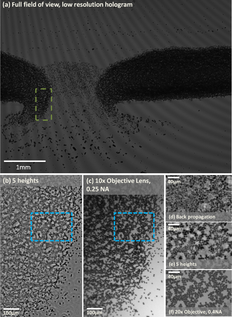
Fig. 3.
(a) Full FOV, LR hologram. (b) Multi-height based PSR lensfree amplitude image of a dense RBC smear is shown. This lensfree image was reconstructed using five different heights (λ = 550nm). The FOV corresponds to the green dashed rectangular in (a). (c) A 10 × objective lens (0.25NA) microscope image is provided for comparison. (d) A single height back propagated PSR amplitude image. The image FOV corresponds to the dashed blue rectangular in (b) and (c). (e) Multi-height based PSR lensfree amplitude image acquired using five different heights is shown. This FOV corresponds to the same FOV as in (d). (f) A 20 × objective lens (0.4 NA) microscope image is also provided for comparison purposes.

