Fig. 4.
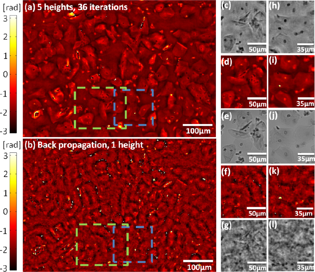
(a) Multi-height based PSR lensfree phase image of a Pap test is shown. This image was reconstructed using five heights. 36 iterations were used during phase recovery (λ = 550nm). (b) Single height back propagated PSR phase image is shown. (c) and (d) are multi-height based PSR lensfree amplitude and phase images, respectively, of the green dashed rectangle shown in (a). The absorbing nuclei of the cells are clearly visible in the amplitude images, while the cell’s boundaries are more visible in the phase image. The corresponding 40× (0.65NA) microscope image is provided for comparison in (e). (f) and (g) are the corresponding single height based back propagated phase and amplitude images respectively. (h) and (i) are multi-height based PSR lensfree amplitude and phase images, respectively, of the blue dashed rectangle in (a). The corresponding 40 × (0.65NA) microscope image is also provided for comparison in (j). (k) and (l) are the corresponding single height based back propagated phase and amplitude images respectively. All the phase images in the figure are wrapped since we did not employ phase unwrapping algorithms.

