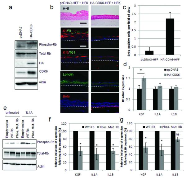
Figure 4. Rb inactivation enhances KGF expression.
A) HA tagged CDK6 expression in HFFs confirmed by Western blotting. B) Organotypic cultures grown with HA CDK6 expressing fibroblasts were stained for differentiation markers and Brdu incorporation which is quantified in C). D) Q PCR analysis of KGF, IL1A and IL1B in HA CDK6 expressing HFFs. * p<0.05 in a Students T test. E) Western blot detection of wild type Rb (WT Rb) and a non phosphorylatable mutant (Phos.Mut.Rb) expressed in HFFs. F) Q PCR assessment of the induction of KGF, IL1A and IL1B expression following 10 ng/mL IL1A (F), or IL1B (G) treatment of cells described in E. Induction of KGF/IL1A and IL1B expression in WT Rb HFFs was assigned as 100%, results are from triplicate experiments. * represents p<0.05 in a Students T test compared to WT Rb samples.

