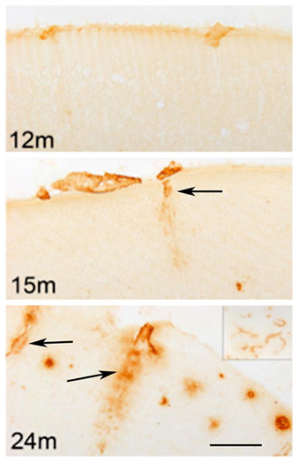
Fig. 3.

The progression of cerebral amyloid angiopathy pathology in amyloid precursor protein Tg2576 mice at 12, 15 and 24 months of age (12, 15, and 24 m). Immunostaining is performed using an antibody specific for Aβ40, which is the predominant amyloid peptide deposited in cerebral amyloid angiopathy. Parenchyma-penetrating blood vessels with amyloid angiopathy are noted by arrows. (Scale bar: 100 μm). A lower magnification image (4×) of frontal cortex with extensive cerebral amyloid angiopathy is shown in the top right corner of 24-month-old mouse
