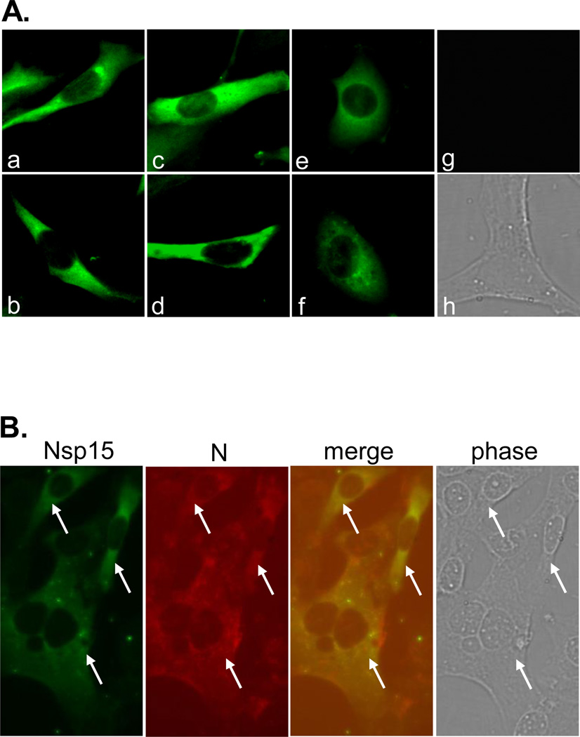
Fig. 1. Nsp15 expression in MHV-A59 infected cells.
(A) 17Cl-1 cells were infected with MHV-A59 at an m.o.i. of 5. At 7 h p.i., nsp15 protein expression was detected with immunofluorescence staining using rabbit anti-nsp15 antibody D23 and goat anti-rabbit IgG-FITC. Panels a-f show the various subcellular localizations of nsp15 in infected cells. Mock-infected cells were used as a negative control (g for fluorescence staining & h for phase contrast). (B) Dual immunofluorescence staining. Infected cells were stained with D23 and monoclonal antibody J3.3 to MHV N protein and detected with anti-rabbit IgG-FITC (Nsp15, green) and anti-mouse IgG-TRITC (N, red). The two colors are then super-imposed (merge) and the phase contrast image (phase) shows both infected and uninfected cells in the same field. The white arrows highlight that all nsp15-expressing cells (green) are virus-infected cells (red). Note that the exposure time for Panels A and B was different.

