Fig. 2.
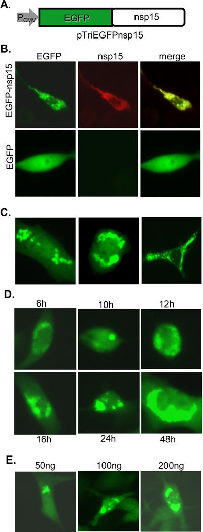
(A) Diagram of expression plasmid MHVEGFPnsp15 showing the MHV nsp15 coding sequence fused at the N-terminus to EGFP. (B) Expression of MHV EGFP-nsp15 or EGFP alone following plasmid transfection. Left panels indicate direct detection of EGFP (EGFP) while middle panels show the detection of nsp15 following immunofluorescence staining with anti-nsp15 antibody D23 and anti-rabbit IgG-TRITC (red). Color-merged images are shown on the right (merge). (C) Examples of detailed speckle formation at various subcellular localizations following the expression of MHV EGFP-nsp15. (D) Time course experiment showing speckle formation from 6 to 48 h post transfection with MHVEGFPnsp15. (E) Speckle formation at 24 h posttransfection with MHVEGFPnsp15 DNA at various concentrations (50 to 200 ng).

