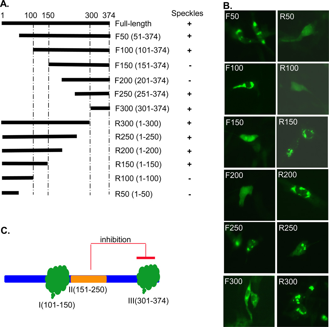
Fig. 4.
Schematic diagram, name and amino acid position of MHV nsp15 deletion mutants used in the experiments in reference to the full-length nsp15. These deleted fragments were fused with EGFP as fusion proteins as in Fig. 2A. (B) Expression and distribution of EGFP-nsp15 deletion mutants. A summary of the ability of individual fusion proteins to form speckles is shown on the right to the corresponding construct in panel (A). (C) Diagram illustrating the domain mapping results, highlighting 3 potentially separate domains (Domain I: aa101–150, Domain II: aa151–250, and Domain III:aa301–374) that regulate speckle formation.

