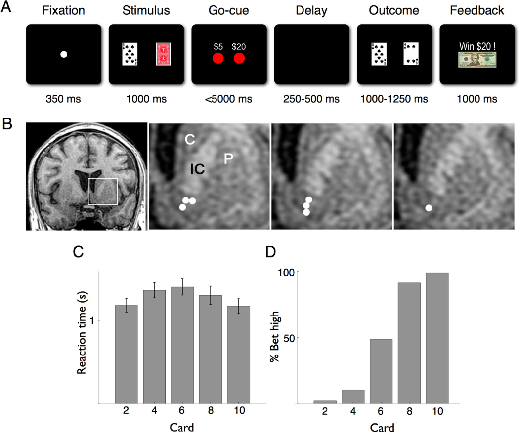
Fig 1.
Behavioral task, recording location, and subject performance. (A) The Fixation screen was followed by the Stimulus screen showing the subject’s card and the back of the opponent’s card. Appearance of the wager choices ($5 or $20) signified the Go-Cue. After a variable delay period, the Outcome screen showed both the subject’s and revealed opponent’s card. The Feedback screen then explicitly displayed how much was won or lost, or whether the hand was a draw. (B) Coronal T1-weighted MRI showing the recording sites from the seven subjects in whom individual neurons were isolated. The first panel shows a slice 2 mm anterior to the anterior commissure (AC). The following three panels show the inset area (left striatum) 2, 3, and 4 mm anterior to the AC, respectively, with recording sites in the ventral striatum denoted by white circles. C, caudate; P, putamen; IC, anterior limb of internal capsule. (C) Average reaction time (in seconds) of all subjects by card value. Error bars are S.E.M. (D) Percentage of high wager ($20) trials by card value.

