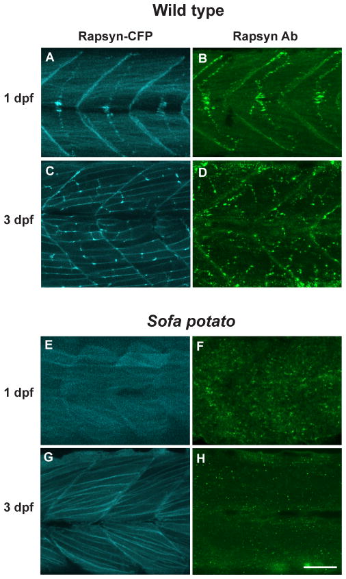
Fig 3.
Rapsyn was visualized in a rapsyn-CFP (+) larva with CFP (A, C and E, G) and in native larva stained with rapsyn-antibody (B, D and F, H). The distribution of CFP and anti-rapsyn antibody staining were similar at 1 dpf (A, B) or 3 dpf (C, D). The distribution of rapsyn was also examined in the sofa potato background, with (E, G) or without (F, H) the rapsyn-CFP transgene. Scale: 50μm.

