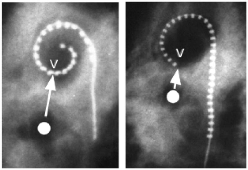
Figure 10.

An anatomical landmark V in our cochlear specimens was defined as the point on the upper basal turn closest to the center of the vestibule. X-ray images here show implanted Contour and Banded arrays with the electrode nearest V indicated. The distance required to reach V (~1000 Hz for both OC and SG) for an electrode positioned under the OC is about 20 mm, while for an ideally positioned perimodiolar array this distance would be only about 12–13 mm in the average cochlea.
