
Figure 1.
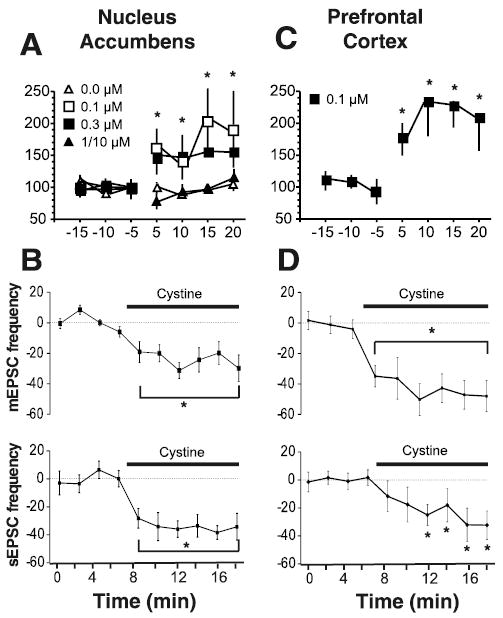
Cystine regulates glutamate levels and excitatory transmission. A, Cystine increased extracellular glutamate in NA slices, as revealed by microdialysis (n = 5 per dose). The 1/10 μm dose is pooled data for perfusion of 1 μm (n = 2) and 10 μm (n = 3). Data were normalized to the average of the three baseline samples and are shown as mean ± SEM percentage change. Basal levels of glutamate (picomoles per sample) are as follows: 0 = 0.176 ± 0.023; 0.1 = 0.134 ± 0.016; 0.3 = 0.125 ± 0.019; 1/10 = 0.136 ± 0.040. B, Cystine (0.3 μm) reduced mEPSC (n = 11; 0.9 ±0.2 Hz) and sEPSC (n = 9 baseline; 0.8 ±0.2 Hz) frequency in medium spiny cells (Vm = −80 mV) in the NA. Data are presented as a percentage change from baseline ±SEM. C, Cystine (0.1 μm) increased extracellular glutamate in prefrontal cortex slices (n = 6). Data were normalized to percentage change from baseline; the basal level was 0.084 ± 0.015. D, Cystine (0.1 μm) reduced mEPSC (n = 12, 2.3 ± 0.6 Hz) and sEPSC (n = 8; 4.9 ±0.8 Hz) frequency in PFC slices. *p <0.05, comparing cystine treatment to the average baseline value using a one-way ANOVA with repeated measures and least significant difference post hoc comparison.