
Figure 3.
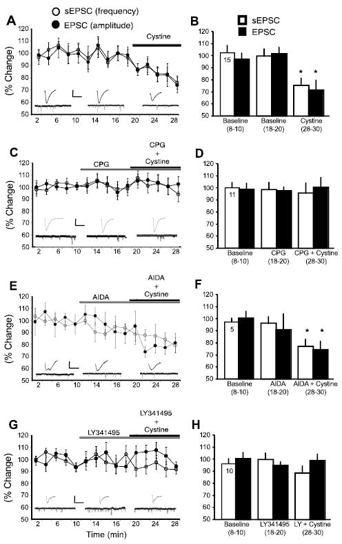
xc- and mGluR2/3 regulate excitatory transmission in the NA. A, C, E, G, Data were normalized to the average of baseline and are shown as mean ± SEM percentage change. Data in B, D, F, and H were collapsed during 8 –10, 18 –20, and 28 –30 min of each experiment and compared with baseline (n is shown in the first bar). A, B, Cystine (0.3 μm) reduced sEPSC frequency and evoked EPSC amplitude after 20 min of baseline. Evoked EPSC traces from a single cell (calibration: 300 pA, 10 ms) and 1 s current traces from the same cell during aforementioned treatments are shown. C, D, The xc- antagonist CPG (1 μm) prevented cystine (0.3 μm) from reducing sEPSC frequency and EPSC amplitude. Evoked EPSC traces from a single cell (calibration: 200 pA, 10 ms) and 1 s current traces from the same cell during aforementioned treatments are shown. E, F, Cystine (0.3 μm) reduces sEPSC frequency and EPSC amplitude in the presence of the group I metabotropic glutamate receptor antagonist AIDA (1 mm). Evoked EPSC traces from a single cell (calibration: 100 pA, 10 ms) and 1 s current traces from the same cell during aforementioned treatments are shown. G, H, The mGluR2/3 antagonist LY341495 (LY) (0.3 μm) prevented cystine (0.3 μm) from reducing sEPSC frequency and EPSC amplitude. Evoked EPSC traces from a single cell (calibration: 100 pA, 10 ms) and 1 s current traces from the same cell during aforementioned treatments are shown. *p <0.05, comparing cystine treatment with the average baseline value using a one-way ANOVA with repeated measures and least significant difference post hoc comparison.