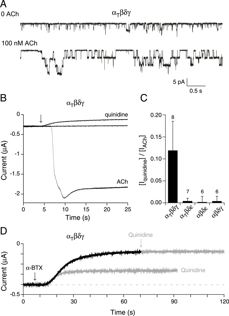
Figure 7. Openings by non-liganded αtwiβδγ receptors expressed in Xenopus oocytes.
A, Outside-out patch recordings of αtwiβδγ receptors before (top trace) and after (bottom trace) addition of ACh (n= 6 patches). B, Recordings of macroscopic current in oocytes expressing αtwiβδγ and held at −50 mV. Shown are the baseline current in the absence of ACh, the decrease in holding current upon addition of 100 µM quinidine (arrow) and, following wash out of the quinidine, the large inward current that occurred following addition of 300 nM ACh (arrow) to the oocyte. C, Overall comparisons of the effect of quinidine to reduce the holding current in the absence of ACh activation of the different receptor isoforms. The fractional peak current was determined on the basis of reduction in holding current in the presence of 100 µM quinidine relative to total ACh activated current following washout of quinidine. The mean ± SD are shown and the number of oocytes tested is indicated. (D) Traces from an oocyte expressing αtwiβδγ receptors showed decreases in holding current in response to individual application of either 1 µM α-btx (black trace) or 100µM quinidine. The response to the first application of quinidine was allowed to recover before the oocyte was treated with α-btx. Following treatment with α-btx the oocyte showed no response to further application of quinidine (grey arrow).

